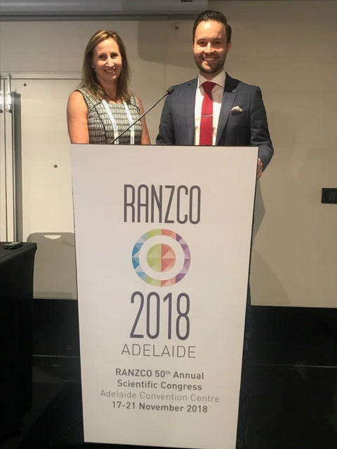
In November 2018 I was invited by ZEISS to present at the RANZCO Annual Scientific Congress in Adelaide. The presentation related to a program called FORUM which enables patient imaging to be imported into practice management software with greater efficiency.
Since the installation of this program our Tasmanian Eye Institute research students have been using FORUM to extract raw data from our OCT imaging across multiple timepoints (patient appointments) to investigate the progression of geographic atrophy. The advanced form of age-related macular degeneration (ARMD) is characterised by the presence of geographic atrophy or exudative changes secondary to choroidal neovacularisation (CNV). Geographic atrophy (GA) is defined as atrophic areas with discrete retinal depigmentation at least 175um in diameter with a sharp border, and visible underlying choroidal vessels (Brader et al. 2011). However GA and CNV do not represent independent pathways for ARMD progression, often occurring in conjunction. Eyes with GA may develop CNV, altering its natural history and creating uncertainty regarding the cause of vision loss. Similarly, vascular endothelial growth factor (VEGF) has been implicated in paracrine and autocrine regulatory pathways of choriocapillaris and RPE, and thus blockade of VEGF pathways has been postulated to play a role in the progression of GA noted in patients treated for CNV (Grisanti & Tatar. 2008). This study aims to investigate the development and progression of geographic atrophy (GA) in patients undergoing intravitreal therapy (IVT) for treatment of choroidal neovascularization (CNV) secondary to age related macular degeneration (ARMD).
Jennie Rossetto and Penny Allen

